Category: Uncategorized

  • Ebola disease

    Key facts

    • Ebola disease is a severe, often fatal illness in humans.
    • Three different viruses are known to cause large Ebola disease outbreaks: Ebola virus, Sudan virus and Bundibugyo virus.
    • The average Ebola disease case fatality rate is around 50%. Case fatality rates have varied from 25–90% in past outbreaks.
    • Early intensive supportive care with rehydration and the treatment of symptoms improves survival.
    • Approved vaccines and treatments are only available for one of the viruses (Ebola virus) and are under development for the others.
    • Outbreak control relies on a package of interventions including intensive supportive care of patients, infection prevention and control, disease surveillance and contact tracing, laboratory services, safe and dignified burials, vaccination if relevant, and social mobilization.

    Overview

    Ebola disease (EBOD) is a rare but severe illness in humans (1). It is often fatal.

    Ebola disease is caused by viruses that belong to the Orthoebolavirus genus of the filoviridae family (2). Six species of Orthoebolaviruses have been identified to date, with three known to cause large outbreaks:

    • Ebola virus (EBOV) causing Ebola virus disease (EVD)
    • Sudan virus (SUDV) causing Sudan virus disease (SVD)
    • Bundibugyo virus (BDBV) causing Bundibugyo virus disease (BVD).  

    Ebola disease first occurred in 1976 in two simultaneous outbreaks: one outbreak was of Sudan virus disease in Nzara in what is now South Sudan, and the other outbreak was of Ebola virus disease in Yambuku, in what is now the Democratic Republic of the Congo. The latter occurred in a village near the Ebola River, from which the disease takes its name.

    While there are licensed vaccines and therapeutics for Ebola virus disease, there is no approved vaccine or treatment for other Ebola diseases, such as SVD or BVD. Candidate products are in development.

    Early intensive supportive care including rehydration and treatment of specific symptoms, can improve survival. Seeking early care can be lifesaving.
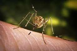
    Transmission

    It is thought that fruit bats of the Pteropodidae family are natural hosts of the Orthoebolavirus. The virus can get into the human population when people have close contact with the blood, secretions, organs or other bodily fluids of infected animals such as fruit bats, chimpanzees, gorillas, monkeys, forest antelope or porcupines found ill or dead or in the rainforest.

    People can get infected with the virus from another person by direct contact (through broken skin or mucous membranes) with:

    • the blood or body fluids of a person who is sick with or has died from Ebola disease; and
    • objects or surfaces that have been contaminated with body fluids (like blood, feces, vomit) from a person sick with the disease or who has died from the disease.

    People cannot transmit the disease before they have symptoms, and they remain infectious as long as their blood contains the virus.

    Health and care workers have frequently been infected while treating patients with Ebola disease. This occurs through close contact with patients when infection control precautions are not strictly practiced.

    Burial ceremonies that involve direct contact with the body of a person who has died can also contribute to the transmission of Ebola disease.
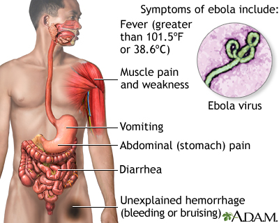
    Symptoms

    The incubation period or interval from infection to onset of symptoms varies from 2 to 21 days.

    The symptoms of Ebola disease can be sudden and include fever, fatigue, malaise, muscle pain, headache and sore throat. These are followed by vomiting, diarrhoea, abdominal pain rash, and symptoms of impaired kidney and liver functions. It is important for health and care workers to be on the lookout for these symptoms.

    Despite a perception that bleeding is a common symptom, this is less frequent and can occur later in the disease. Some patients may develop internal and external bleeding, including blood in vomit and faeces, bleeding from the nose, gums and vagina. Bleeding at the sites where needles have punctured the skin can also occur.

    The impact on the central nervous system can result in confusion, irritability and aggression.

    Diagnosis

    It can be difficult to clinically distinguish Ebola disease from other infectious diseases such as malaria, typhoid fever, shigellosis, meningitis and other viral haemorrhagic fevers because symptoms at early stage of the disease are similar.

    Confirmation that the person has an Orthoebolavirus infection is made using the following diagnostic methods:

    • reverse transcriptase polymerase chain reaction (RT-PCR) assay
    • antibody-capture enzyme-linked immunosorbent assay (ELISA)
    • antigen-capture detection tests
    • virus isolation by cell culture.

    Samples collected from patients are an extreme biohazard risk; laboratory testing on non-inactivated samples should be conducted under maximum biological containment conditions. All non-inactivated biological specimens should be packaged using the triple packaging system when transported nationally and internationally See Diagnostic testing for Ebola and Marburg diseases.

    Treatment

    Over the years, WHO and partners have developed guidance and training that outline how to provide the best possible care for patients and increase their chance of survival, whether or not specific treatments are being used. Called optimized supportive care, this covers the relevant tests to administer, how to manage pain, nutrition and co-infections (such as malaria), and other approaches that put the patient on the best path to recovery.

    For Ebola virus disease, WHO made strong recommendations for treatment with mAb114 (ansuvimabTM) or REGN-EB3 (InmazebTM) that are both monoclonal antibodies. For other Ebola diseases, such as SVD or BVD, there are no approved therapeutics, but candidate products are under development and a CORE protocol for clinical trials is available.

    Vaccines

    For Ebola virus disease:

    For other Ebola diseases, such as SVD:

    Prevention and control

    Community engagement is key to successfully controlling any outbreak. Outbreak control relies on using a range of interventions, such as clinical care, surveillance and contact tracing, laboratory services, infection prevention and control in health facilities, safe and dignified burials, vaccination (only for Ebola virus disease) and social mobilization.

    Raising awareness of risk factors and protective measures that individuals can take is an effective way to reduce human transmission. Risk reduction messaging should focus on several factors:

    • Reduce the risk of wildlife-to-human transmission from contact with infected fruit bats or monkeys/apes and the consumption of their raw meat.
    • Reduce the risk of human-to-human transmission arising from direct or close contact with infected people, particularly with their body fluids. Close physical contact with Ebola patients should be avoided. Patients should be isolated in a designated treatment center for early care and to avoid transmission at home.
    • Communities should be well informed, both about the disease itself and how to control the outbreak. This is done best when they are involved in the response and there is open discussion.
    • Outbreak containment measures include safe and dignified burial of the deceased, identifying people who may have been in contact with someone infected with Ebola disease and monitoring their health for 21 days, separating the healthy from the sick to prevent further spread and providing care to confirmed patients. Maintaining good hygiene and a clean environment are also important.

    Controlling infection in health-care settings

    Health-care workers should always take standard precautions when caring for patients, regardless of their presumed diagnosis. These include basic hand hygiene, respiratory hygiene, use of personal protective equipment (to block splashes or other contact with infected materials), safe injection practices and safe and dignified burial practices.

    Health-care workers caring for patients with suspected or confirmed Ebola disease should apply extra infection control measures to prevent contact with the patients’ blood and body fluids and contaminated surfaces or materials such as clothing and bedding. Infection prevention and control guideline for Ebola and Marburg diseases.

    Laboratory workers are also at risk. Samples taken from humans and animals for investigation of Orthoebolavirus infection should be handled by trained staff and processed in suitably equipped laboratories.

    Care for survivors

    All survivors, their partners and families should be shown respect, dignity and compassion. WHO does not recommend isolation of recovered patients whose blood has tested negative for Orthoebolavirus. Survivors might suffer from both clinical and psychological sequelae. WHO encourages affected countries to consider the establishment of care programme to alleviate sequelae, support to community reintegration, counselling and biological testing.

    Orthoebolaviruses are known to persist in immune-privileged sites in some people who have recovered. These sites include the testicles, the inside of the eye and the brain. Relapse-symptomatic illness in the absence of re-infection in someone who has recovered from Ebola disease is rare but has been documented. Reasons for this phenomenon are not yet fully understood.

    Ebola virus transmission via infected semen has been documented up to fifteen months after clinical recovery. To mitigate the risk of this transmission, a semen testing programme should be implemented to:

    • offer counselling to male survivors and their sexual partners to inform them of the potential risk and support them adhering to safer sex practices (including condom provision and good hand and personal hygiene);
    • offer monthly semen testing until they have had two consecutive negative test results; and
    • after two consecutive negative tests, survivors can safely resume normal sexual practices with minimized risk of virus transmission.

    In the absence of a semen testing programme, male survivors should follow safer sex practices for 12 months.

    Orthoebolavirus may persist in the placenta, amniotic fluid and fetus of women infected while pregnant, and in the breast milk of breastfeeding women who are infected with the virus. Survivor care programmes should encompass care for pregnant and breastfeeding women after their recovery.  

    WHO response

    WHO works with countries to prevent Ebola outbreaks by maintaining surveillance and supporting at-risk countries to develop preparedness plans. The following document provides overall guidance for control of Ebola and Marburg virus outbreaks: Ebola and Marburg virus disease epidemics: preparedness, alert, control, and evaluation

    When an outbreak is detected, WHO responds by supporting outbreak response, disease detection, community engagement, contact tracing, vaccination, vaccine and treatment trials, case management, laboratory services, infection control, logistics, and training and assistance with safe and dignified burial practices.

  • Japanese encephalitis

    Key facts

    • Japanese encephalitis virus (JEV) is a flavivirus related to dengue, yellow fever and West Nile viruses, and is spread by mosquitoes (especially Culex tritaeniorhynchus).
    • JEV is the main cause of viral encephalitis in many countries of Asia with an estimated 100 000 clinical cases every year (1).
    • Although symptomatic Japanese encephalitis (JE) is rare, the case-fatality rate among those with encephalitis can be as high as 30%. Permanent neurologic, cognitive and behavioural sequelae occur in 30–50% of those with encephalitis.
    • The majority of cases occur in children below 15 years of age.
    • Twenty-four countries in the WHO South-East Asia and Western Pacific Regions have endemic JEV transmission, exposing more than 3 billion people to risks of infection.
    • There is no cure for the disease. Treatment is focused on relieving severe clinical signs and supporting the patient to overcome the infection.
    • Safe and effective vaccines are available to prevent JE. WHO recommends that JE vaccination be integrated into national immunization schedules in all areas where JE disease is recognized as a public health issue.

    Overview

    Japanese encephalitis virus (JEV) is an important cause of viral encephalitis in Asia. It is a mosquito-borne flavivirus and belongs to the same genus as dengue, Zika, yellow fever and West Nile viruses. The first case of Japanese encephalitis viral disease (JE) was documented in 1871 in Japan. The annual incidence of clinical disease varies both across and within endemic countries, ranging from 10 per 100 000 population or higher during outbreaks. A literature review and modelling study estimates about 100 000 clinical cases (95% CI: 61 720–157 522) of JE globally each year, with approximately 25 000 deaths (95% CI: 14 550–46 031). JE primarily affects children. Most adults in endemic countries have natural immunity after childhood infection, but individuals of any age may be affected.

    Signs and symptoms

    Most JEV infections are mild (fever and headache) or without apparent symptoms, but approximately 1 in 250 infections results in severe clinical illness. The incubation period is 4–14 days. In children, gastrointestinal pain and vomiting may be the dominant initial symptoms. Severe disease is characterized by rapid onset of high fever, headache, neck stiffness, disorientation, coma, seizures, spastic paralysis and ultimately death. The case fatality rate can be as high as 30% among those with disease symptoms. Of those who survive, 20–30% suffer permanent cognitive, behavioural or neurological sequelae such as seizures,  hearing or vision loss, speech, language, memory, and communication problems or weakness of the limbs.

    Transmission

    Twenty-four countries in the WHO South-East Asia and Western Pacific Regions have JEV transmission risk, which includes more than 3 billion people. JEV is transmitted to humans through bites from infected mosquitoes of the Culex species (mainly Culex tritaeniorhynchus). Humans, once infected, do not develop sufficient viraemia to infect feeding mosquitoes. The virus exists in a transmission cycle between mosquitoes, pigs and/or water birds (enzootic cycle). The disease is predominantly found in rural and periurban settings, where humans live in closer proximity to these vertebrate hosts, in particular domestic pigs. In most temperate areas of Asia, JEV is transmitted mainly during the warm season, when large epidemics can occur. In the tropics and subtropics, transmission can occur year-round but often intensifies during the rainy season and pre-harvest period in rice-cultivating regions.

    Diagnosis

    Individuals who live in or have travelled to a JE-endemic area and experience encephalitis are considered a suspected JE case. Initial diagnosis of JE can be made by clinical examination followed by a lumbar puncture. A laboratory test is required to confirm JEV infection and to rule out other causes of encephalitis. WHO recommends testing for JEV-specific IgM antibody in a single sample of cerebrospinal fluid (CSF) or serum, using an IgM-capture ELISA. If tested negative, a convalescent sample may be tested. Testing of CSF sample is preferred to reduce false-positivity rates from previous infection or vaccination.

    Surveillance of the disease is mostly syndromic for acute encephalitis syndrome. Confirmatory laboratory testing is often conducted in dedicated sentinel sites, and efforts are undertaken to expand laboratory-based surveillance. Case-based surveillance is established in countries that effectively control JE through vaccination.

    Treatment

    Encephalitis is a medical emergency and requires urgent medical attention. There is no antiviral treatment for patients with JE. Treatment is supportive and includes stabilization and relief of  symptoms.  

    Those who have lived through encephalitis often have health-care needs requiring long-term treatment and care including rehabilitation. The ongoing psychosocial impacts of disability from encephalitis can have medical, educational, social and human rights-based implications. Despite the high burden of sequelae on people with encephalitis, their families and the community, access to both services and support for these conditions is often insufficient, especially in low- and middle-income countries. Individuals and families with members disabled by encephalitis should be encouraged to seek services and guidance from local and national Organizations of Disabled People (ODPs) and other disability focused organizations, which can provide vital advice about legal rights, economic opportunities and social engagement to ensure people disabled by encephalitis are able to live full and rewarding lives.

    Prevention and control

    Progress has been made in Asia with the implementation of JE vaccination programmes, with most endemic countries having country-wide or targeted programmes in place.  A decline in incidence of the disease has been reported in recent years, which is likely due in part to vaccination. Gavi supports JE catch-up campaigns and co-finances the vaccine for routine immunization in eligible countries.

    Safe and effective JE vaccines are available to prevent disease. WHO recommends having strong JE prevention and control activities, including JE immunization in all regions where the disease is a recognized public health priority, along with strengthening surveillance and reporting mechanisms. Even if the number of JE-confirmed cases is low, vaccination should be considered where there is a suitable environment for JE virus transmission. Introduction of the vaccine should be done in conjunction with a one-time catch-up campaign.

    There are three main types of JE vaccines currently in use: several inactivated Vero cell-derived vaccines, a live attenuated vaccine, and a live recombinant (chimeric) vaccine. One inactivated vaccine and the two live vaccines are WHO prequalified.

    The risk to travellers to Japanese encephalitis-endemic areas is normally low, but travellers should take precautions to avoid mosquito bites. Personal preventive measures include the use of mosquito repellents, long-sleeved clothes, coils and vaporizers. Travellers spending extensive time in JE endemic areas are recommended to get vaccinated before travel.

    In endemic areas, there is little evidence to support a reduction in JE disease burden from interventions other than the vaccination of humans. Thus, vaccination of humans should be prioritized over vaccination of pigs and mosquito control measures. However, the spread of JEV in new areas has been correlated with agricultural development and intensive rice cultivation supported by irrigation programmes.

    WHO response

    WHO responds to Japanese encephalitis in the following way:

    • supports countries in the confirmation of outbreaks through its collaborating network of laboratories;
    • develops surveillance standards and case definitions for reporting;
    • provides guidance on clinical management of disease and long-term care;
    • supports vector control efforts in conjunction with the Global Vector Control Response;
    • develops guidance on the optimal use of vaccines through the publication of vaccine position papers;
    • prequalifies vaccines as a service to UNICEF and Gavi.
    • WHO is implementing the Intersectoral global action plan on epilepsy and other neurological disorders in consultation with Member States to address many challenges and gaps in providing care and services for people with epilepsy and other neurological disorders such as JE that exist worldwide.
  • Dengue fever

    Key facts

    • Dengue is a viral infection caused by the dengue virus (DENV), which is transmitted to humans through the bite of infected mosquitoes.
    • About half of the world’s population is now at risk of dengue, with an estimated 100–400 million infections occurring each year.
    • Dengue is found in tropical and sub-tropical climates worldwide, mostly in urban and semi-urban areas.
    • While many DENV infections are asymptomatic or produce only mild illness, DENV can occasionally cause more severe cases, and even death.
    • Prevention and control of dengue rely on vector control. There is no specific treatment for dengue/severe dengue, and early detection and access to proper medical care greatly lower fatality rates of severe dengue.

    Overview

    Dengue (break-bone fever) is a viral infection that is spread from mosquitoes to people. It is more common in tropical and subtropical than in temperate climates.

    Most people who get dengue do not have symptoms. For those who do, the most common symptoms are high fever, headache, body aches, nausea and rash. Most get better in 1–2 weeks. Some develop severe dengue and need care in a hospital. 

    In severe cases, dengue can be fatal.  

    You can lower your risk of dengue by avoiding mosquito bites, especially during the day.

    Dengue is treated through pain management as there is no specific treatment currently.

    Symptoms

    Most people with dengue have mild or no symptoms and will get better in 1–2 weeks. Rarely, dengue can be severe and lead to death.  

    If symptoms occur, they usually begin 4–10 days after infection and last for 2–7 days. Symptoms may include:

    • high fever (40°C/104°F)
    • severe headache
    • pain behind the eyes
    • muscle and joint pains
    • nausea
    • vomiting
    • swollen glands
    • rash. 

    Individuals who are infected for the second time are at greater risk of severe dengue. The symptoms of severe dengue often come after the fever has gone away and may include:

    • severe abdominal pain
    • persistent vomiting
    • rapid breathing
    • bleeding gums or nose 
    • fatigue
    • restlessness
    • blood in vomit or stool
    • being very thirsty
    • pale and cold skin
    • feeling weak.

    People with these severe symptoms should seek care immediately.  

    After recovery, people who have had dengue may experience fatigue for several weeks.

    Diagnostics and treatment

    Laboratory-based and point of care diagnostics are critical to control and manage dengue, yet global disparities in laboratory capabilities present significant challenges. The diagnostic algorithms, testing strategies and test methodologies employed vary, depending on the capabilities of national laboratory systems. The wide range of available tests – including nucleic acid amplification tests (NAATs), enzyme-linked immunosorbent assays (ELISAs) and rapid diagnostic tests (RDTs) –  vary significantly in quality and performance.

    Laboratory testing for arboviruses can be accomplished through either direct detection methods such as virus isolation, molecular detection of nucleic acid or antigen testing, including rapid diagnostic tests (RDTs) within the first week of illness.

    There is no specific treatment for dengue, although pain can be managed with medication such as paracetamol (acetaminophen). Non-steroidal anti-inflammatory medicines such as ibuprofen and aspirin should be avoided as they can increase the risk of bleeding.

    For people with severe dengue, hospitalization is often necessary.

    Global burden

    The incidence of dengue has grown dramatically worldwide in recent decades, with the number of cases reported to WHO increasing from 505 430 cases in 2000 to 14.6 million in 2024. The vast majority of cases are asymptomatic or mild and self-managed, and hence the actual numbers of dengue cases are under-reported. The disease is now endemic in more than 100 countries.

    In 2024, more cases of dengue were recorded than ever before in a 12-month period, affecting over 100 countries on all continents. During 2024, ongoing transmission, combined with an unexpected spike in dengue cases, resulted in a historic high of over 14.6 million cases and more than 12 000 dengue-related deaths reported. The Region of the Americas contributed a significant proportion of the global burden, with over 13 million cases reported to WHO. 

    Several factors are associated with the increasing risk of spread of the dengue epidemic, including the changing distribution of the responsible vectors (chiefly Aedes aegypti and Aedes albopictus), especially in previously dengue-naive countries; climate change leading to increasing temperatures, high rainfall and humidity; fragile and overburdened health systems; limitations in surveillance and reporting; and political and financial instabilities in countries facing complex humanitarian crises and high population movements.

    One modelling estimate indicates 390 million dengue virus infections per year, of which 96 million manifest clinically(1).A recent study on the prevalence of dengue estimates that 5.6 billion people are at risk of infection with dengue and other arboviruses(2).From January to July 2025, over 4 million cases and over 3000 deaths have been reported to WHO from 97 countries.

    Dengue is spreading to new areas, including the European and Eastern Mediterranean regions. In 2024, 308 cases were reported to WHO from three European countries (France, Italy and Spain) and an additional 1291 cases and four deaths were recorded in the French overseas territories of Mayotte and Réunion. 

    Transmission

    Transmission through the mosquito bite

    The dengue virus is transmitted to humans through the bites of infected female mosquitoes, primarily the Aedes aegypti mosquito. Other species within the Aedes genus can also act as vectors, but their contribution is normally secondary to Aedes aegypti.

    After feeding on a DENV-infected person, the virus replicates in the mosquito midgut before disseminating to secondary tissues, including the salivary glands. The time it takes from ingesting the virus to actual transmission to a new host is termed the extrinsic incubation period (EIP). The EIP takes about 8–12 days when the ambient temperature is 25–28°C. Variations in the EIP are not only influenced by ambient temperature but also by several other factors – such as the magnitude of daily temperature fluctuations, the virus genotype, and the initial viral concentration – which can also alter the time it takes for a mosquito to transmit the virus. Once infectious, a mosquito can transmit the virus for the rest of its life.

    Human-to-mosquito transmission

    Mosquitoes can become infected by people who are viremic with DENV. This can be someone who has a symptomatic dengue infection, someone who is yet to have a symptomatic infection (those who are pre-symptomatic), and also someone who shows no signs of illness (those who are asymptomatic).

    Human-to-mosquito transmission can occur up to 2 days before someone shows symptoms of the illness, and up to 2 days after the fever has resolved.

    The risk of mosquito infection is positively associated with high viremia and high fever in the patient; conversely, high levels of DENV-specific antibodies are associated with a decreased risk of mosquito infection. Most people are viremic for about 4–5 days, but viremia can last as long as 12 days.

    Maternal transmission

    The primary mode of transmission of the DENV between humans involves mosquito vectors. There is evidence, however, of the possibility of maternal transmission (i.e. from a pregnant mother to her baby). At the same time, vertical transmission rates appear low, with the risk of vertical transmission seemingly linked to the timing of acquiring the dengue infection during pregnancy. When a mother does have a dengue infection when she is pregnant, babies may suffer from pre-term birth, low birthweight and fetal distress.

    Other transmission modes

    Rare cases of transmission via blood products, organ donation and transfusions have been recorded. Similarly, transovarial transmission of the virus within mosquitoes has also been recorded. 

    Risk factors

    Previous infection with DENV increases the risk of an individual developing severe dengue.

    Urbanization (especially rapid, unplanned urbanization), is associated with dengue transmission through multiple social and environmental factors: population density, human mobility, access to reliable water source, water storage practices, etc.

    Community risks to dengue also depend on population knowledge, attitudes and practices towards dengue, as exposure is closely related to behaviours such as water storage, plant-keeping and self-protection against mosquito bites. Routine vector surveillance and control activities and targeted community engagement greatly enhance resilience. 

    Vectors can adapt to new environments and climate. The interaction between the dengue virus, the host and the environment is dynamic. Consequently, disease risks may change and shift with climate change in tropical and subtropical areas, in combination with increased urbanization and population movement.

    Prevention and control

    The mosquitoes that spread dengue are active during the day. 

    To lower your risk of getting dengue, protect yourself from mosquito bites by using: 

    • clothes that cover as much of your body as possible;
    • mosquito nets, ideally sprayed with insect repellent, if sleeping during the day;
    • window screens;
    • mosquito repellents (containing DEET, Picaridin or IR3535); and
    • coils and vaporizers.

    To prevent mosquitoes from breeding:

    • implement environmental management and modification practices to stop mosquitoes from accessing egg-laying habitats;
    • dispose of solid waste properly and remove artificial habitats that can hold water;
    • cover, empty and clean domestic water storage containers on a weekly basis; and
    • apply appropriate insecticides to water storage outdoor containers.

    If you get dengue, it’s important to:

    • rest;
    • drink plenty of liquids;
    • use acetaminophen (paracetamol) for pain;
    • avoid non-steroidal anti-inflammatory medication such as ibuprofen and aspirin; and
    • watch for severe symptoms and contact your doctor as soon as possible if you notice any.

    Currently, one vaccine (QDenga) is available and licensed in some countries. However, it is recommended only for those aged 6–16 years in high transmission settings. Several additional vaccines are under evaluation.

    WHO response

    WHO responds to dengue by:

    • supporting countries in the confirmation of outbreaks through its collaborating network of laboratories;
    • providing technical advice and guidance to countries for the effective management of dengue outbreaks;
    • supporting countries to improve their reporting systems and capture the true burden of the disease;
    • providing training on clinical management, diagnosis and vector control at the country and regional levels in collaboration with its collaborating centres;
    • formulating evidence-based strategies and policies;
    • supporting countries to develop dengue prevention and control strategies and adopt the Global Vector Control Response (2017–2030) and the Global Arbovirus Initiative (2022–2025);
    • reviewing and making recommendations on the development of new tools, including insecticide products and application technologies;
    • gathering official records of dengue and severe dengue from over 100 Member States; and
    • publishing guidance and handbooks for surveillance, case management, diagnosis, dengue prevention and control for Member States.